EMA (Epithelial Membrane Antigen) Antibody (Immunohistochemistry)
The EMA (Epithelial Membrane Antigen) Antibody is one of the most widely used
immunohistochemical markers for identifying epithelial and epithelial-like tumors.
EMA is a transmembrane glycoprotein that is normally expressed in a broad range of epithelial tissues.
As a result, it plays an important role in tumor classification.
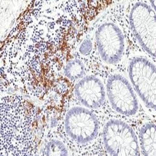
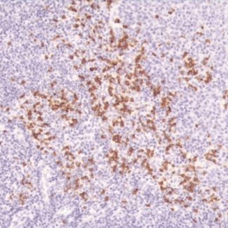
In IHC staining, EMA shows membranous or cytoplasmic positivity in many tumors.
These include breast, ovarian, lung, colon, and renal carcinomas. Moreover, EMA expression is also seen
in mesotheliomas, synovial sarcomas, and plasma cell neoplasms. Therefore, EMA helps pathologists
distinguish epithelial tumors from lymphoma or melanoma.
🔬 Features and Benefits
- Specific marker for epithelial and epithelial-differentiated tumors
- Clear brown membrane or cytoplasmic staining
- Helps differentiate carcinoma from lymphoma and melanoma
- Compatible with standard IHC protocols
- Stable for up to 18 months at 2–8 °C
💡 Diagnostic Applications
- Detection of adenocarcinoma and squamous cell carcinoma
- Identification of malignant mesothelioma
- Differentiation of carcinoma from sarcoma and lymphoma
- Diagnosis of synovial sarcoma and epithelioid sarcoma
- Evaluation of plasma cell neoplasms and Hodgkin lymphoma
🌐 Fardad Azma Rad Co.
Fardad Azma Rad Co., the exclusive representative of
Talent Biomedical in Iran, provides high-quality IHC antibodies including
EMA. The company remains committed to diagnostic accuracy, scientific precision,
and international standards. Therefore, it supports pathology and molecular laboratories across the country
with reliable and advanced products.


There are no reviews yet.